
iStar 334T CCD
View Product
 Part of the Oxford Instruments Group
In Raman Spectroscopy, photons which have been excited in the vibrational levels in a molecule, are scattered so that they gain or lose energy. This inelastic scattering provides information about the vibrational states of the molecule. A simplified energy diagram that illustrates these concepts is shown below.
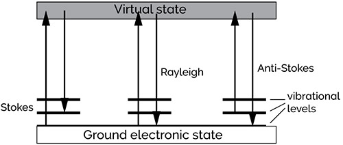
Simplified energy diagram
If no energy change occurs, which happens in most cases, we call it the Rayleigh transition. This is the most intense band in the Raman spectrum, as can be seen in the representation of the spectrum below.
Stokes radiation occurs at lower energy (longer wavelength) than the Rayleigh radiation, and anti-Stokes radiation has greater energy. The energy increase or decrease is related to the vibrational energy levels in the ground electronic state of the molecule. The observed Raman shift of the Stokes and anti-Stokes features is a direct measure of the vibrational energies of the molecule. A schematic Raman spectrum may appear as shown below.
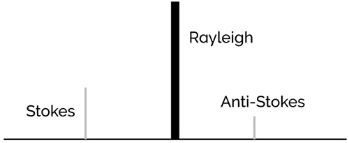
Schematic Raman spectrum
The energy of the scattered radiation is less than the incident radiation for the Stokes line and the energy of the scattered radiation is more than the incident radiation for the anti-Stokes line. The energy increase or decrease from the excitation is related to the vibrational energy spacing in the ground electronic state of the molecule and therefore the wavenumbers of the Stokes and anti-Stokes lines are a direct measure of the vibrational energies of the molecule.
In the example spectrum, notice that the Stokes and anti-Stokes lines are equally displaced from the Rayleigh line. This occurs because in either case one vibrational quantum of energy is gained or lost. Also, note that the anti-Stokes line is much less intense than the Stokes line. This occurs because only molecules that are vibrationally excited prior to irradiation can give rise to the anti-Stokes line. Hence, in Raman spectroscopy, only the more intense Stokes line is normally measured. Raman scattering is a relatively weak process - the number of photons Raman scattered is quite small. However, there are several processes which can be used to enhance the sensitivity of a Raman measurement.
The Resonance Raman effect
If the wavelength of the exciting laser coincides with an electronic transition of a molecule, the intensity of Raman-active vibrations from the chromophore are enhanced by a factor of 102 to 104. This resonance enhancement or resonance Raman effect can be extremely useful, not just in significantly lowering the detection limits, but also in introducing electronic selectivity. Thus, the resonance Raman technique is used to provide both structural and electronic information across a range of biological and chemical species.

Metalloporphyrins, carotenoids and several other classes of biologically important molecules have strongly allowed electronic transitions in the visible, making them ideal candidates for resonance Raman spectroscopy. The spectrum of the chromophore is resonance enhanced and that of the surrounding environment is not. For biological chromophores, this means that absorbing active centres can be specifically probed by visible excitation wavelengths, and not the surrounding protein matrix (which would require UV lasers to bring it into resonance).
Resonance Raman spectroscopy is also an important probe of the chemistry of metal-centred complexes, fullerenes, polydiacetylenes and other "exotic" molecules which strongly absorb in the visible. Although many more molecules absorb in the ultraviolet, the high cost of lasers and optics for this spectral region have limited ultraviolet (UV) resonance Raman spectroscopy to a small number of specialist groups.
Vibrations which are resonantly enhanced fall into two or three general mechanistic classes. The most common case is Franck-Condon enhancement. In this, a component of the normal coordinate of the vibration occurs in a direction in which the molecule expands during an electronic excitation. The more the molecule expands along this axis when it absorbs light, the larger the enhancement factor. The easily visualized ring breathing (in-plane expansion) modes of porphyrins fall into this class.
Vibrations which couple two electronic excited states are also resonantly enhanced, through a mechanism called vibronic enhancement. In both cases, enhancement factors roughly follow the intensities of the absorption spectrum.
Resonance enhancement does not begin at a sharply defined wavelength. In fact, enhancement of 5x to 10x is observed if the exciting laser is within even a few hundred wavenumbers below the electronic transition of a molecule. This "pre-resonance" enhancement can be experimentally useful.
Techniques and Applications Using Raman Spectroscopy
Here are four examples of techniques using Raman Spectrosopy
1. SERS – Surface-Enhanced Raman Scattering
The Raman scattering from a compound (or ion) adsorbed on or even within a few Angstroms of a structured metal surface can be 103 to 106 times greater than in solution. This surface-enhanced Raman scattering (SERS) is strongest on silver but is observable on gold and copper as well. At practical excitation wavelengths, enhancement on other metals is unimportant.
SERS arises from two mechanisms:
Other aromatic nitrogen or oxygen containing compounds, such as aromatic amines or phenols, are strongly SERS active. The effect can also be seen with other electron-rich functionalities such as carboxylic acids.
The intensity of the surface plasmon resonance is dependent on many factors including the wavelength of the incident light and the morphology of the metal surface. The best morphology for surface plasmon resonance excitation is a small (<100 nm) particle or an atomically rough surface. SERS is commonly used to study monolayers of materials adsorbed on metals, including electrodes. Other surfaces that have been studied are colloids and metal films on dielectric substrates. SERS is not generally used for analytical purposes due to non-reproducibility of measurements.
2. UVRRS - Ultra Violet Resonance Raman Spectroscopy
UVRRS is a powerful tool in the molecular analysis of complex biological systems including proteins and DNA. Most biological systems absorb UV radiation and hence have the ability to offer resonance with UV Raman excitation. For example, excitation around 200 nm enhances the Raman peaks from vibrations of amide groups; excitation around 220 nm enhances peaks from certain aromatic residues. The Raman scatter from water is weak, allowing for analysis of very weak aqueous systems.
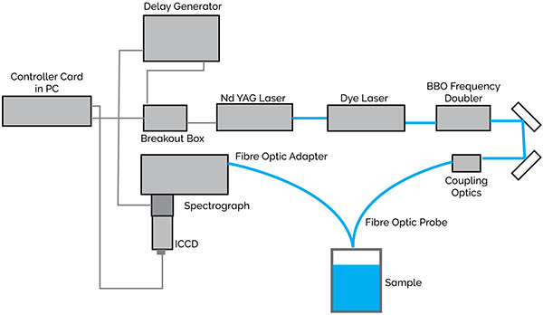
Fibre optic UVRRS configuration
A tunable laser is required as the excitation source so that the resonant wavelengths can be selected. An Nd:YAG-pumped dye laser with frequency-doubled output is suitable. Depending on the dyes used, this laser setup can give almost any required UV wavelength. Intensified CCDs (ICCDs) with UV photocathodes, back-illuminated CCDs or CCDs with UV enhancing (BASF lumogen) coatings can be used as detectors for UVRRS. These detectors are used on account of their high detection efficiency and multichannel capabilities.
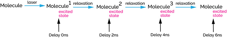
Pulsed lasers can be used to study short-lived excited states. A laser pulse can be supplied to a molecular system with enough energy to redistribute the electrons in a molecule causing the formation of an excited state as illustrated below.
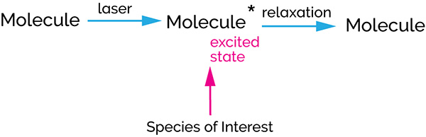
Species of Interest
The Raman spectrum of this excited state molecule can be studied either using the same laser pulse or a different pulse from a second laser (single colour and two-colour pulsed Raman). Excited states can have lifetimes, from picoseconds to milliseconds, but the majority can be studied using a gating in the order of 5 ns. As the majority of excited states are generated using UV and visible lasers, photocathodes with high UV and visible Quantum Efficiencies (QEs) are typically suitable.
Schematic of pump-probe (two color) Raman
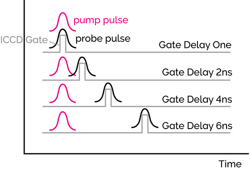
Time delay sequences
The simplest pulsed laser experiments are single-color experiments where high irradiance laser pulses are used both to initiate the photoreaction, and then to Raman probe the excited state. By opening an intensifier tube, only the Raman spectrum of the excited state will be recorded. This pulse/ICCD gate combination will be repeated and accumulated hundreds to thousands of times in order to achieve a good overall signal-to-noise ratio with high dynamic range.
3. TR3 Time Resolved Resonance Raman Spectroscopy
In Time Resolved Resonance Raman (TR3) spectroscopy, pairs of laser pulses of different wavelength are used to photolyse (optically pump) and then to Raman probe the transient species of interest. The spectral window of the spectrograph/detector is chosen so that it corresponds to the frequency range of the Raman scattering from the probe laser.
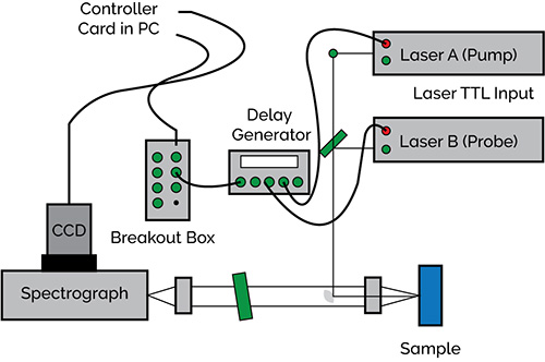
Pulsed two color Raman layout with delays under the control of a delay generator
The time dependence of the transient signal is monitored by recording a series of spectra at different delays after the photolysis, i.e. at a series of time delays between the excitation and probe pulses. The ICCD camera or either of the lasers can supply the trigger. A delay generator is used to control the delays.
4. Raman Microscopy
In Raman microscopy, a research grade optical microscope is coupled to the excitation laser and the spectrometer. This produces images down to 1 micron and generates Raman Spectra. Imaging and spectroscopy can be combined to generate Raman cubes which are three-dimensional data sets, yielding spectral information at every pixel of the 2D image.
If a motorized xyz microscope stage is used to automatically record spectral files, Raman maps can be recorded. Specific software routines will allow the quick and easy reconstruction of these maps.
© Oxford Instruments 2024